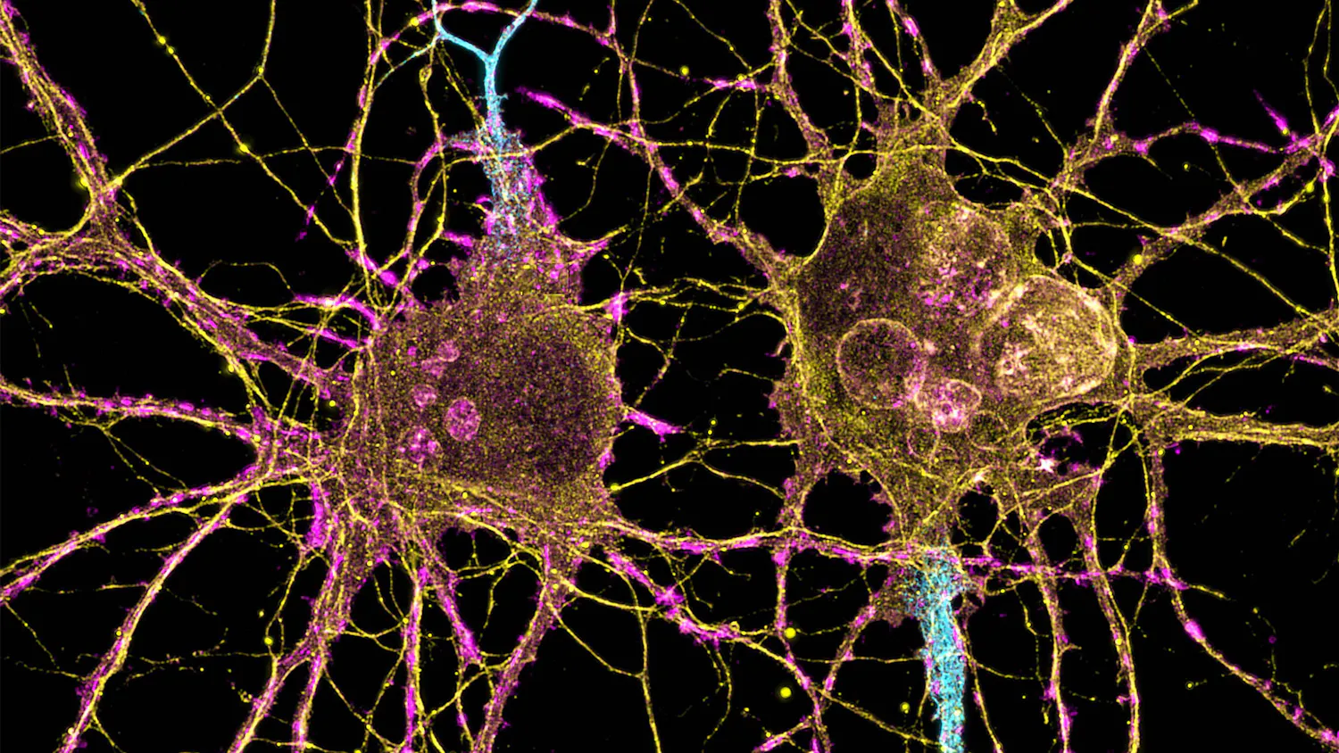
Collagen is the most abundant protein in the human body, making up a huge amount of our bone, muscle and tendon mass. Collagen plays a key role in tissue function and stability. It is also a key indicator of tissue health. For these reasons, identifying the presence of collagen and noting its intrinsic structure can prove valuable in the healthcare field.
Traditional optical microscopes are a crucial part of medical research. The problem? Collagen, at a molecular level, is completely transparent. Researchers here at UW-Madison are working around that.
A group in the Department of Biomedical Engineering led by Paul Campagnola has created a novel solution to this molecule’s inconvenient attribute. “In some ways it’s a simple idea but no one has actually done it before,” Campagnola said, referring to his group’s new machine which includes a 3-D printed component that holds a rotating motor and a sample collection tube to an upright microscope.
With this device, collagen can now be imaged with clarity. It is a key step toward the future of integrating personalized medicine into patient care for a variety of illnesses.
Collagen takes on different structures depending on the health of the tissue it is in. In healthy tissue, it appears in a uniform grid-like pattern. If the tissue is cancerous, the collagen is distorted into different formations. Because of the complicated nature of cancer, “the only commonality of all cancers is that the collagen is remodeled,” Campagnola said, “It’s remodeled in all epithelial cancers. The things you think of as potentially cancerous: organs, skin, everything… it all has collagen remodeling.”
Once applied to a clinical setting, the ability to view cancer patient collagen can better inform healthcare professionals and patients alike on the state of the illness and how to best move forward with treatment.
Imaging collagen on a patient-specific level is one of the many innovations being categorized as personalized medicine. Personalized medicine is the tailoring of specific treatments based on a patient’s unique condition.
According to Campagnola, the collagen imaging technique will improve healthcare for patients of many different conditions aside from cancer. “It’s for cancer, but a lot of other diseases as well. Cardiovascular diseases, autoimmune diseases, there’s connective tissue disorders, fibroses… but we are primarily working on cancer and pulmonary fibrosis.”
All of these diseases can be detected and classified by the state of collagen tissue and it has been impossible to assess collagen tissue structure without an accurate method for viewing it until now.
However, a feat like this did not come overnight. This is why Campagnola dedicated almost 20 years to the project.
Before his arrival at UW-Madison in 2010, Campagnola was at the University of Connecticut Health Center, where he worked with traditional microscopic imaging. “[Traditional imaging] allows you to take individual 2-D sections, so you can image one plane at a time, then you can take a series of them and build them up into three dimensions,” Campagnola said.
Unfortunately, collagen is completely visible only when the molecule is perpendicular to the plane of sight. Campagnola takes a pen and holds it in such a way that the whole profile of the utensil is visible, ball-point to barrel. When the pen is held so that you’re facing directly at the point of it, barely any of it is visible. This barely visible perspective is how collagen is configured within a three-dimensional matrix of tissue.
“Because the [microscope] has the property that your sample is mounted on the desk, the laser is coming in [facing the point]… you don’t see the collagen molecules. And then as you tilt them closer and closer to the plane, they will get brighter.”
Collagen at this parallel angle is invisible, unmeasurable and essentially not quantifiable. Moreover, because if its 3-D nature, no matter what angle you look at it, a substantial amount of the tissue will always be hidden.
How is collagen imaging done then? A tissue sample is placed into the new machine that completely rotates the sample while taking images at every angle. “We took our tissue and we put it in a tube so it’s just like this pen and we spun it so we get views at all different angles. We take a three-dimensional data set at each angle. So if we have a fiber that’s transparent, when we rotate it in this way, we can see [the fiber].”
Once the images are taken at every single angle, they are combined into one 3-D model using a complicated set of algorithms. This multidisciplinary project required collaboration from many departments. “There was engineering in making the mechanical structure to do this, but the equal challenge is the mathematical reconstruction where you take all of these images at all these different angles and put them back together. That’s a very serious challenge.”
“We’re going to improve our experimental aspects so we can get better resolution, but also we are going to improve our reconstruction algorithms. We’re starting those things now.” Campagnola added.
“We have something that works, but it’s nowhere near optimal,” Campagnola admitted. “We have demonstrated proof of principle, but it may not be good enough for what we really want to do. We actually have a lot of work to do.”
Campagnola predicts that this technology will be implemented into a healthcare setting in just five to ten years from now.
This technology is not only a possible diagnostic tool for cancer and other collagen-related disease, but also a possible method of understanding countless other structures of the human body. Many other molecules have visibility as obscure as collagen. Machines such as the one that Campagnola helped develop can enhance our understanding of the human body and what optimal health looks like at a molecular level.